
Introduction
This experiment aimed to identify antibodies against the Human Immunodeficiency Virus (HIV) in simulated patient samples using the enzyme-linked immunosorbent test (ELISA). The virus known as HIV targets and kills immune system CD4 cells, which results in the development of acquired immune deficiency syndrome (AIDS). The ELISA test detects antibodies against HIV antigens, indicating exposure to the virus, by utilizing the body’s unique immune response to pathogens. As to the hypothesis, people who have been directly exposed to HIV sources or who are at high risk of doing so will test positive, whereas people who have had little to no exposure will test negative. This theory is based on the core ideas of immunology, which state that antigens trigger specific immune responses and that antibody production serves as a defensive mechanism.
Materials/Methods
Materials
In the orchestrated setting of our ELISA simulation, an array of meticulously selected materials created an environment reflective of the assay’s traditional application for detecting antibodies against the Human Immunodeficiency Virus (HIV). At the foundation of this experimental setup were the microtiter plates, which were specifically pre-coated with synthetic HIV antigens to reflect the virus’s antigenic complexity. Segmented into numerous wells, these plates facilitated the simultaneous analysis of various samples, setting the stage for a robust comparative study. The simulated antigen solution was central to demonstrating the antibody-antigen interaction and pivotal to the ELISA detection mechanism. This synthetic concoction emulated the HIV antigens, ensuring the experiment’s fidelity to the principles underlying antibody-mediated immune responses.
The control solutions further defined the experimental framework, dividing it into positive and negative contingencies. Equipped with antibodies ready to bind the HIV antigen, the positive control was designed to yield a predictable positive result, serving as a harbinger of the assay’s operational success. By comparison, the negative control lacked these antibodies, providing a baseline against which to assess and validate the specificity of the observed reactions. The group of patient samples with the labels A through F represented a range of potential HIV exposure situations. These samples were essential in evaluating the ELISA’s diagnostic proficiency across various viral contact scenarios, from direct exposure to minimal risk.
The assay’s detection strategy relied on observing antigen-antibody complexes, enabled by a simulated secondary antibody conjugated to an enzyme. This combination effectively linked the physical, observable markers of antibody presence with the molecular details of the immune response. The chromogen substrate, a colorimetric agent, acted on this conjugate, causing a color change due to its interaction with the enzyme.
The transition from an inert to a colorful state indicated the presence of HIV-specific antibodies in the samples. It provided insight into how the participants’ exposure to HIV affected their serological makeup. By using this carefully designed interaction of artificial materials, the ELISA simulation went beyond its didactic premise and embodied the assay’s fundamental function in viral diagnostics.
Methods
The ELISA simulation technique was carefully crafted to mimic the steps of a conventional indirect ELISA while introducing the essential simplifications appropriate for a learning environment. The experiment started with carefully prepping microtiter plates. Three drops of the simulated HIV antigen solution were added to each well in rows A and B. This procedure was carried out as precisely as possible, using separate plastic pipettes for each sample to eliminate the risk of cross-contamination. This is evidence of the strict standards maintained even in a simulated setting.
After carefully positioning the antigen, the experiment added patient samples and control solutions to the appropriate wells. Three drops of the appropriate solution were placed in each of the wells marked A1-A3 for the positive control and A4-A6 for the negative control. The distribution of the patient samples, labeled A through F, into the subsequent wells, carefully adding each sample in triplicate, came next. This methodology aimed to enhance the reliability of the collected data by ensuring replicable results and mitigating any disparities arising from pipetting errors.
The next critical step was adding the simulated secondary antibody to each well, signaling the transition from the primary antigen-antibody interaction to the assay’s visual result. Here, three carefully timed drops of the secondary antibody were added, initiating the detection process that powers the ELISA. Applying the chromogen substrate, a crucial interaction that caused a color change inside the wells, brought the experiment to a successful conclusion. The conjugated secondary antibody’s enzymatic activity on the substrate caused this color shift, a reliable predictor of whether HIV-specific antibodies were present in the samples.
The microtiter plate was incubated at room temperature for five to ten minutes during the final phase to allow the colorimetric response to develop fully. After this incubation period, the wells were examined closely, and the resulting color changes were carefully noted, giving concrete proof of the experiment’s results. While simplified for educational purposes, this experimental design retained the essential components of the ELISA method, demonstrating its critical function in identifying specific antibodies and, consequently, in diagnosing illnesses. Notably, the decision to omit the washing steps that are usually essential to an extensive ELISA protocol was deliberate to streamline the simulation process, demonstrating a careful balancing act between learning goals and the complex realities of laboratory procedures.
Results
An analytical summary of the data was made possible by methodically organizing the ELISA simulation’s experimental results into a table (see Table 1), which displays each sample’s subsequent color changes and their corresponding test results. The positive control wells in this organized display showed a dark purple color, indicating that the assay can produce a positive result when tested against recognized HIV antibodies. Bursa et al. stressed the importance of accuracy and reliability in interpreting ELISA results, and this result provided crucial validation of the ELISA setup’s operational integrity, confirming its diagnostic precision.
Table 1: ELISA Simulation Results for HIV Antibody Detection
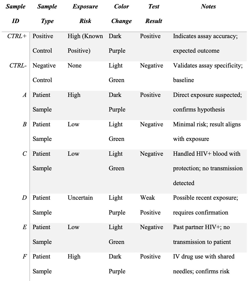
On the other hand, the negative control wells stubbornly retained their initial light green hue, indicating a poor outcome. This sharp difference in the control findings confirmed the assay’s specificity and provided a standard against which to compare the patient samples’ results. Interestingly, patient samples A and F exhibited a strong presence of HIV antibodies, supporting a positive infection status, by mirroring the dark purple coloration of the positive control wells. The ELISA’s capacity to detect seropositivity across various exposure settings is highlighted by its alignment with the positive control, reflecting the ELISA’s sophisticated understanding of antibody-antigen interactions, which is essential to its diagnostic methodology.
Patient D’s sample produced a light purple color with a subtle deviation, indicating a weakly positive result. This specific hue, distinct from the baseline light green but less intense than the unmistakable dark purple, suggests a complex antibody profile that may indicate early infection or a lower antibody concentration. These results demonstrate the ELISA’s sensitivity to changing antibody levels.
These results align with Ambrosioni et al.’s emphasis on the importance of timely and precise diagnostics in managing infectious diseases. The lack of color change in samples from patients B, C, and E, mirroring the negative control, indicates the absence of HIV antibodies, confirming their negative status. This underscores the ELISA’s specificity in identifying non-infected cases, particularly in lower-risk scenarios, and reflects core principles of the immune response.
These findings show a direct relationship between the ELISA test results and the estimated exposure risks suggested for each patient. The congruence of these findings with the original theories underscores the diagnostic validity of the ELISA and its promise as a vital tool for managing and preventing infectious diseases. This experiment connects the theoretical understanding of antibody-mediated immunity with the real-world applications of serological testing, as discussed in the seminal studies by Bursa et al. and Ambrosioni et al., by adhering to the procedural nuances of ELISA and conducting a reflective analysis of patient samples.
Discussion
The assay’s ability to identify antibodies against HIV is thoroughly examined in the discussion of the ELISA simulation results, which supports the theory of the relationship between test results and exposure risk. Positive results were observed for Patients A and F, consistent with the expected reaction in direct or high-risk exposure settings. This agreement highlights the fundamental idea behind ELISA: the use of the immune response to pathogens. This idea is further supported by the careful work of Ambrosioni et al., who explain the subtleties of immune system activation in the presence of viral pathogens such as HIV.
An exciting result is shown in the instance of Patient D: a faint positive result suggesting possible recent HIV exposure. The assay’s sensitivity to different antibody concentrations in the sample is highlighted by the assay’s mild response, which may indicate an early infection stage where antibody concentrations have not yet peaked. Bursa et al. emphasize that fluctuations in antibody levels can affect the interpretation of ELISA assays, underscoring the importance of accuracy in ELISA investigations.
Therefore, the faintly positive result demonstrates the importance of the early HIV detection window, where antibody levels are detectable but not conclusive, and verifies the ELISA’s diagnostic sensitivity. This result is consistent with the larger conversation, as highlighted by Ambrosioni et al., about the promptness of HIV diagnosis and its consequences for patient care and treatment plans.
Conversely, the unfavorable results for Patients B, C, and E demonstrate the assay’s specificity, which successfully differentiates between exposure risks and the ensuing serological signs. These negative results further demonstrate the robustness of ELISA in accurately capturing serostatus following the postulated risk exposure, as they align with the lower risk profiles of the corresponding patients. This specificity is important in the larger context of HIV diagnostics because clinical decision-making and patient counseling depend on a clear distinction between infected and non-infected persons.
Despite its educational value, the simulation adds a layer of abstraction from clinical realities by diverging from the standard ELISA methodology, including removing wash steps and using simulated reagents. Although these changes make educational goals clearer, they also highlight the complexity and rigor of diagnostic processes. Bursa et al. emphasize that the subtle dynamics of ELISA, from sample preparation to the last detection phase, highlight the painstaking attention to detail needed in clinical diagnostics. Therefore, although this deviation is useful for modeling, it requires careful interpretation of the data when considering the assay’s clinical application.
Additionally, Ambrosioni et al. have highlighted the importance of confirmatory testing in HIV diagnoses, which is further highlighted by the appearance of a weak positive result. Because serological assays are inherently complex and can yield results that range from positive to negative, this requirement arises. In clinical practice, indeterminate results necessitate using a second, more decisive testing method to determine the person’s serostatus. Ensuring diagnosis accuracy, guiding patient management methods, and reducing the psychological effects of unknown HIV test findings all depend on this stage.
Conclusion
The ELISA simulation experiment successfully illustrated the assay’s theory and its use in identifying anti-HIV antibodies that reflect individual viral exposure. Positive outcomes were correlated with established risk factors for HIV transmission, hence providing support for the theory. This experiment emphasizes the significance of ELISA in diagnosing viral infections and the vital role the immune system plays in identifying and treating disease. Uncertain cases require further investigation and thorough testing, highlighting the ongoing need for advances in the biomedical sciences to improve disease detection and preventive measures.
Works Cited
Ambrosioni, Juan, et al. “Overview of SARS-CoV-2 Infection in Adults Living with HIV.” The Lancet HIV, vol. 8, no. 5, 2021.
Bursa, Francis, et al. “Estimation of ELISA Results Using a Parallel Curve Analysis.” Journal of Immunological Methods, vol. 486, 2020.
